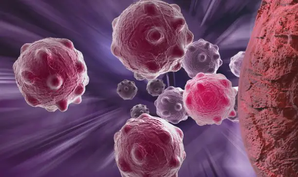
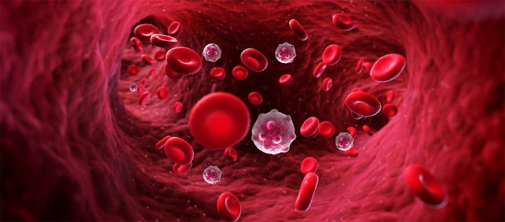
Being able to detect the circulating cancer cells (CTC) in the blood of cancer patients is a topic of great interest to medical oncologists. The promise of be able to detect such rare cells consistently and reproducibly has attracted considerable attention in recent years. However, the rarity of CTCs made this promise difficult to deliver.

On a very basic level, CTCs are cancerous cells that have been cleared from a primary tumor or metastatic site, which circulate through blood and / or lymphatic vessels as single cells or aggregates. A subset of CTCs survive, decant into interstitial space, and eventually form a tumor in a new and distant microenvironment.
CTC analysis offers a unique opportunity for the development of new biomarkers and the characterization and monitoring of the progression of metastatic disease, which can provide key information for new clinical treatments. For these reasons, preclinical metastasis models are increasingly used to study CTCs and are also established directly using CTCs isolated from patient blood samples or after expansion. ex vivo.
CTCs were identified in mice using patient-derived xenograft (PDX) models from breast cancer. This type of model provides a constant and renewable source of CTC, allowing the study of CTCs and the underlying metastatic processes.
Breast cancer CTC cell lines have also been established and used to identify a potential brain metastasis signature. CTCs with this signature were highly invasive and generated brain and lung metastases when xenografted into immunocompromised mice.
Similarly, CTCs for breast cancer were isolated from patients with estrogen receptor positive breast cancer and used to establish cell lines, 60% of which were carcinogenic in mice.
Restricted circulating CTCs required enrichment of blood cells prior to detection and, too often, such enrichment leads to further loss of CTC, making them even rarer.
A diagnostic study at the Carole & Ray Neag Comprehensive Cancer Center at theUConn Health led by Dr. Susan Tannenbaum, head ofHematology-Oncology and head of the service Neag Comprehensive Cancer Center and Dr. Emily Hsu, member ofHematology-Oncology Fellowship Program, is evaluating CTCs in breast cancer patients using a new microscope platform for the detection of circulating cells developed by QCDx LLC (Quantitative Cell Diagnostix), a medical device company located at theUConn Technology Incubation Program (TIP) to Farmington, Connecticut.
CTC: the new microscope platform manages to track them
RareScope is uniquely designed for identify and characterize CTCs from blood samples without prior enrichment. Thereby, this technology detects the varied diversity of circulating cells in order to focus treatment, based on biomarkers observed in circulating cells, often different from the cells found in a previous biopsy sample.

The investigation began in July 2020 and accumulated 21 breast cancer patients with both early and late stage disease and 13 healthy volunteers.
“In the ongoing CLINBREAC clinical trial we have now collected information on circulating tumor cells using the new RareScope light sheet fluorescence microscopy system to calculate numbers of all circulating tumor cells present in the breast of patients with neoadjuvant and metastatic cancer at multiple time points “, Tannenbaum said.
“Furthermore – The scientist continued – we were able to quantify the expression of epithelial and mesenchymal markers as well as markers HER-2, ERα And Trop2 by immunofluorescent staining. In our limited data available at this time, we were able to follow the course of the disease with these markers that inform us about the next therapeutic steps. The promising technology has the unique potential to personalize and enable the use of multiple concordant targeted therapies that could have a significant impact on patient outcomes. “

These study results CLINBREAC in progress were presented on December 8 at the 2021 Symposium of the American Association for Cancer Research SABCS to San Antonio, in Texas.
The instrument analyzes complete populations of nucleated cells from the patient’s blood sample using three-dimensional optical tomography with specialized image analysis software, which also involves the‘artificial intelligence.
“Our technology allows the analysis of morphologically intact and / or live cells in immobilized cell suspensions. Without enrichment, each individual cell is visualized and intracellular markers are characterized. Rare cells such as CTCs can be detected alongside subpopulations of blood cells including lymphocytes, which can be critically important for optimizing the treatment”.

“In addition, the technology is available to identify target cells that can be isolated for molecular analysis of single cells downstream “, said Dr. Triantafyllos Tafas, founder and CEO of QCDx.
Tafas concluded by saying: “We believe our technology is unique in enabling cellular mapping of genomic, transcriptomic and proteomic disease heterogeneity when tissue biopsy is not possible and can critically support clinicians to improve treatment decisions and benefit outcomes and quality of the life of the patient. We are very happy to develop our technology in collaboration with UConn’s Comprehensive Cancer Center and supported by the UConn Technology Incubation Program“.
#Circulating #tumor #cells #CTCs #technology #tracks


