
A group of researchers from the Massachusetts Institute of Technology (MIT) has developed a new platform that allows study entire hemispheres of the human brain in 3D with unprecedented resolution. This technology offers detailed observation starting from the architecture of the tissue and the morphology of the cells, down to the smallest cellular and molecular details, such as the connections between neurons and expressed proteins.
Their work, published in the journal Science, has already provided new insights into the lesions caused by Alzheimer’s. The ultimate goal is to create a three-dimensional atlas of human brain cells at subcellular resolution, thereby improving understanding of human organ functions and disease mechanisms. This, in turn, could stimulate the development of new therapies.
The three innovative technologies underlying the discovery of the human brain
The MIT team combined three cutting-edge technologies to achieve these results:
- MEGAtome: a vibrating microtome that slices tissue with an ultra-precise cut, keeping the connections between cells intact.
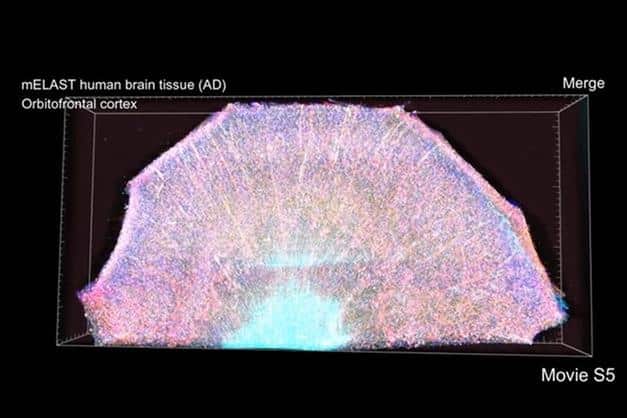
- mELAST: a hydrogel that makes tissue samples clear, elastic, expandable and reversibly markable, allowing studies at multiple scales.
- Unslice: software that reassembles tissue slices to reconstruct the cerebral hemisphere in 3D, restoring the alignment of individual blood vessels and neuronal connections.
A leap forward for medical research
This technological platform not only avoids tissue degradation, but makes them durable and can be analyzed several times over time. Observe entire hemispheres of the human brain this way is a fundamental step for two main reasons. First of all, it allows us to study multiple aspects of a single brain, avoiding variations between the brains of different individuals. Second, the speed and scalability of this method allow for the creation of representative samples of various genders, ages and pathological conditions, facilitating more robust statistical comparisons.
First applications and future of research
There platform it has already been tested on two human brains donated to science: one healthy and one affected by Alzheimer’s. Initially, the researchers explored the orbitofrontal cortex, identifying regions with significant neuron loss. Using various markers, they then highlighted the relationships between pathogenic factors and cell types, discovering that synapse loss is most concentrated in areas with direct overlap of amyloid plaques.
This technology promises to revolutionize the study of neurological diseases and could lead to the creation of one fully mapped brain bank at the subcellular level, ready to be re-analysed with new markers depending on future needs.
#revolutionary #zoom #human #brain #detail


