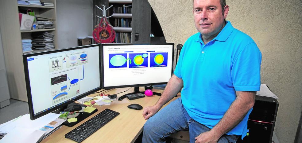
Francisco Cavas, director of the Bioengineering and Applied Computational Simulation Group of the UPCT. /
Researchers from the Polytechnic University of Cartagena use computational modeling and simulation of biological structures applied in medicine

Leaving aside the brain, which some describe as “the most complex and enigmatic structure in the Universe”, we could say that the eye is the most complex organ in the body. In fact, they are closely related because the eyes, by themselves, do not see, but receive light and generate information in the form of neural electrical impulses that reach the brain and that is where they are transformed into images. In short, they are a means of transmission. Although, to do their job well, they have more than 100 million cells and it is one of the most active muscles in the body. In addition, it is made up of different essential structures for the success of its function, such as the lens (responsible for focusing objects located at different distances), the retina (responsible for generating the electrical signals that reach the brain) or the cornea (which covers the iris and the pupil and refracts light), among others.
Given its complexity and the important role that the sense of vision plays in our day-to-day life, there is a great deal of research aimed at improving ophthalmological surgical interventions. At the Polytechnic University of Cartagena, the ‘Bioengineering and Applied Computational Simulation’ group directed by Francisco Cavas dedicates its main line of research to ‘Modelling of Biological Structures’.
“We are dedicated to computational modeling and simulation of biological structures applied in medicine. Specifically applied to the cornea, although we are already working with other structures of interest. In our group we have other lines of research, such as that aimed at medical equipment or the development of new materials for different applications, etc. », he says.
With their models they intend to help clinical decision-making and also to plan surgeries by evaluating their risks.
Regarding the cornea, the team evaluates the efficacy and safety of computational models based on the biomechanical-morphofunctional behavior of the cornea in different scenarios, such as in the detection/diagnosis of diseases or in the evaluation of the biomechanical behavior of the structure after a corrective treatment, whether it is invasive or not, in short, scenarios that occur in clinical practice. With their models they aim to help clinical decision-making, or to help plan surgeries by evaluating their risks, which makes it possible to significantly reduce the extra cost derived from possible post-surgical side effects. In short, says Cavas, “we try to promote of sustainability and efficiency for ophthalmological services in hospitals».
Within the framework of their line of work, they have collaborated, for example, in an investigation with the AIDEMAR association (San Javier) and with Jorge Alió (from the Miguel Hernández University and Vissum Alicante Clinic). Specifically, they have evaluated the singularities of the geometric profiles that cornal architecture presents in children diagnosed with Down syndrome, and their influence on the visual capacity of these patients. The findings obtained allow redefining the current approach on the diagnosis of keratopathy in these patients.
Likewise, in recent years they have managed to patent a procedure and a 3D model to detect keratoconus in its preclinical phase, that is, before the patient shows symptoms. It is a disease whose development in its clinical phase affects the human cornea and involves a progressive loss of visual quality, leading to total loss of vision in its most advanced phase, which makes a corneal transplant necessary. .
They have recently started a collaboration on Fuchs Edema (a corneal disease that occurs when the cells of the innermost layer of the cornea, called the endothelium, progressively die causing reduced vision) with the Ramón y Cajal Hospital in Madrid . They also have other collaborations with ocular prosthesis companies such as the North American ‘AdditonTechnologyInc’, which is the only one ‘approved by the FDA (USA) for the manufacture and marketing of intracorneal rings (used to treat keratoconus).
In short, it is a translational scientific work, which practically reaches the patient immediately. «We have several ongoing collaborations, specifically I would like to highlight the collaboration with the Ophthalmology Service of the Santa Lucía University Hospital in Cartagena, with the Department of Optics of the University of Alicante and with the Department of Pathology and Surgery of the Miguel Hernández University. of Elche within the framework of a research project recently granted to the UPCT by the Carlos III Health Institute of Madrid”, points out Francisco Cavas.
It refers to the project Development and validation of a new concept of biomechanical-morphofunctional characterization of the cornea with which they are working to evaluate the efficacy and safety of several computational models based on the biomechanical behavior of the cornea before and after surgical treatments.
The team led by Cavas stands out for being interdisciplinary, made up of engineers, opticians and ophthalmologists. In this study they intend to create an infrastructure capable of developing a better clinical alternative to the evolutionary diagnosis of keratoconus and a new biomechanical simulation technology capable of intervening in the post-surgical clinical evolution of the cornea. “We believe that this technological advance will represent a paradigm shift in the therapy of debilitating diseases of the cornea, and will allow, through the development of personalized medicine, to improve the health and quality of life related to the visual function of patients.”
#Technology #testing #surgeries