A discovery by Simona Stäger’s team could help find a cure for the most severe form of leishmaniasis. Leishmaniasis is a tropical disease affecting an increasing number of people worldwide. Between 700,000 and 1 million new cases are reported each year.
The research results were published in the journal Cell Reports.
The causes of leishmaniasis
Caused by a parasitic protozoan of the Leishmania genus, which is transmitted to humans through the simple bite of a phlebotomistleishmaniasis includes three clinical forms, of which the visceral form is the most serious.
Professor Stäger from the Institut national de la recherche scientifique (INRS) and her team, in collaboration with other researchers from the INRS and McGill University, have observed a surprising immune mechanism linked to chronic visceral leishmaniasis.
This discovery could be an important step towards a new therapeutic approach to this disease.
In many infections, CD4 T cells play a key role in the defense of the affected organism. Unfortunately, in the case of chronic infections such as leishmaniasis, maintaining the number of functional CD4 cells becomes an important issue, since the immune system is constantly activated to react against the pathogen affecting the infected person.
The study conducted by Professor Stäger in her laboratory at the INRS Armand-Frappier Santé Biotechnology Research Center suggests that these cells may have more than one trick up their sleeves to maintain their viability.
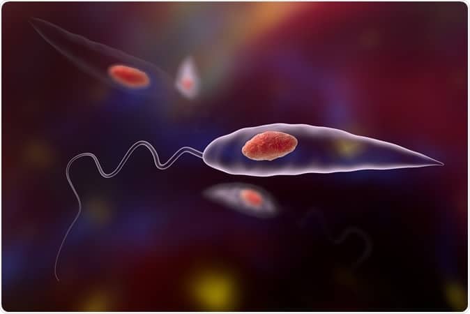
“We discovered a new population of CD4 cells in mice infected with the parasite responsible for visceral leishmaniasis. These T cells have interesting properties,” said Professor Stäger.
Observing these new cells, scientists noticed that they increase in number during the chronic phase of the disease and that, like progenitor cells, they are capable of self-renewal or differentiation into other effector cells responsible for eliminating the parasite, or regulatory cells. cells that inhibit the host response.
Professor Stäger points out that CD4 T cells normally differentiate into effector cells from “naive” CD4 T cells. But during chronic infections, due to the constant need to generate effector cells, naïve CD4 T cells are highly stressed and can become exhausted.
We believe that in the chronic phase of visceral leishmaniasis, the new population we have identified is responsible for the generation of effector and regulatory cells. This would allow the host to prevent depletion of its existing pool of naïve CD4 T cells for a given antigen,” explains PhD student and first author of the study, Sharada Swaminathan.
The new population of lymphocytes discovered by the INRS team could represent a decisive immune enhancer, taking the place of overly stressed naïve CD4 T cells.
“If we could understand how to direct this new population of lymphocytes to differentiate into a protective effector cell, we could help the host get rid of the Leishmania parasite,” said Professor Stäger.
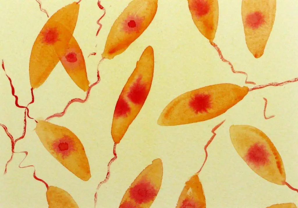
The study also mentions that cells similar to this new population of CD4 T cells were observed in mice infected with the lymphocytic choriomeningitis virus and in mice carrying the intestinal worm H. polygyrus. So it is quite possible that this population is present in other chronic infections or in other chronic inflammatory environments.
This overlap sets the stage for an even broader scope for the discovery made by Professor Stäger’s team. “If our hypothesis is correct, these cells could be exploited at a therapeutic level not only for visceral leishmaniasis, but also for other chronic infections,” concludes the researcher.
This article was co-authored with Sharada Swaminathan, Linh Thuy Mai, Alexandre P. Meli, Liseth Carmona-Pérez, Tania Charpentier, Alain Lamarre, Irah L. King, and Stäger.
Information on a major player in chronic visceral leishmaniasis
In an article appearing in PLOS Pathogens, INRS professor Simona Stäger and her team show how the parasite Leishmania donovani exploits a physiological response to low oxygen levels (hypoxia) to establish a chronic infection.
The visceral leishmaniasis parasite causes chronic inflammation that enlarges the spleen and creates a hypoxic microenvironment. To compensate for the lack of oxygen and ensure their survival, cells adapt by inducing the expression of the transcription factor HIF-1α, the main regulator of the cellular response to hypoxia.
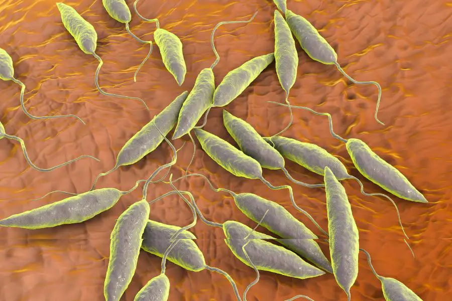
Professor Stäger and her team demonstrated the effect of this key regulator on the function of monocytes and macrophages during viceral leishmaniasis, the most severe form of a tropical disease affecting millions of people worldwide. These cells are the main targets of the L. donovani parasite
“Our laboratory work demonstrates that HIF-1α plays a key role in the establishment of chronic Leishmania infections by reducing the ability of monocytes and macrophages to kill the parasite. We also found that HIF-1α gives these cells immunosuppressive properties,” explains Professor Stäger.
The fight against the Leishmaniasis parasite
Professor Albert Descoteaux’s team from the INRS Center – Institut Armand-Frappier, Canada, has gained a better understanding of how the Leishmania donovani parasite manages to outsmart the human immune system and proliferate with impunity, causing visceral leishmaniasis, a chronic infection potentially fatal if left untreated. This scientific discovery was recently published in PLoS Pathogens.
Approximately 350 million people live in areas where leishmaniasis is possible. Over 90% of cases are reported in India, Bangladesh, Nepal, Sudan and Brazil. Leishmaniasis is also found in Mexico and other parts of South America. There are no effective vaccines to prevent leishmaniasis and resistance problems significantly reduce the effectiveness of conventional drugs.
The parasite, which is transmitted to humans during the blood meal of infected sandflies, is internalized via macrophages in the liver, spleen and bone marrow. However, this parasite manages to alter the normal process of phagocytosis (destruction of foreign bodies), resist this process, replicate and infect other macrophages.
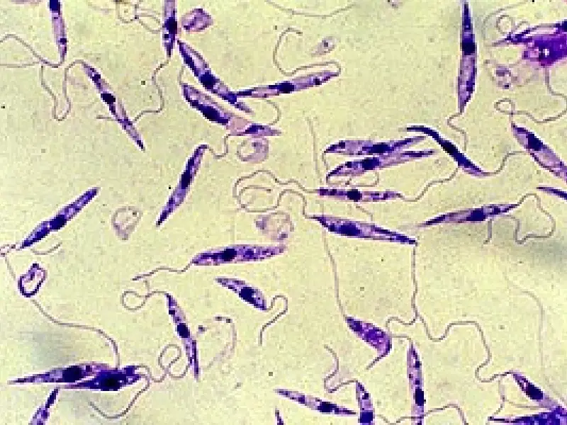
This resistance process involves in particular blocking the normal acidification process within the macrophage by interrupting membrane fusions.
To date, few studies have attempted to identify the regulators of these membrane fusions and their role in the process of phagolysosomal biogenesis (a compartment in which pathogenic microorganisms are usually killed).
The work of PhD student Adrien Vinet and Professor Descoteaux has shed new light on the biology of Leishmania parasites, particularly the molecular mechanisms through which they manage to outsmart the human immune system.
Leishmaniasis parasites escape death by exploiting the immune response to sandfly bites
Cutaneous leishmaniasis, a disease characterized by painful skin ulcers, occurs when the parasite Leishmania major, or a related species, is transmitted to a mammalian host through the bite of an infected sandfly. In a new study from the National Institute of Allergy and Infectious Diseases (NIAID), part of the National Institutes of Health, scientists found that L. major does its damage not only by dodging but also by exploiting the wound-healing response to sand bites of fly.
“This work changes the textbook picture of the life cycle of the leishmaniasis parasite by identifying the inflammatory cell known as the neutrophil as the predominant cell involved during the initiation of infection,” says NIAID Director Anthony S. Fauci, MD
Using advanced microscopic techniques, which enabled real-time imaging of the skin of live mice infected with L. major, NIAID collaborators Nathan C. Peters, Ph.D., and Jackson Egen, Ph.D., discovered that neutrophils, the white blood cells that ingest and destroy bacteria play a surprising role in the development of the disease.
Neutrophils were rapidly recruited from the circulating blood and into the skin of infected mice, where they swarmed around the sandfly bite sites and effectively engulfed the parasites.
Unlike many other infectious organisms that die inside neutrophils, L. major parasites appear to have evolved to escape death, actually surviving for long periods of time inside neutrophils.
The parasites eventually escape from the neutrophils and enter macrophages, another population of immune cells in the skin, where they can establish a long-term infection.
“Parasites transmitted by sandflies to mice lacking neutrophils have more difficulty establishing an infection and surviving. This demonstrates the importance of neutrophils at the site of an infected sandfly bite and suggests the unexpected route taken by the parasite from sandfly to neutrophils to macrophages. it is a critical component of this disease,” says Dr. Peters.
Additionally, Dr. Egen says, the study reveals how neutrophils leave locally inflamed blood vessels and move into tissues; provides new information on the movement of these immune cells within damaged tissue environments and upon contact with pathogens; and provides video images that reveal the active entry of neutrophils into areas of damaged skin.
#Leishmaniasis #hope #cure #forms
