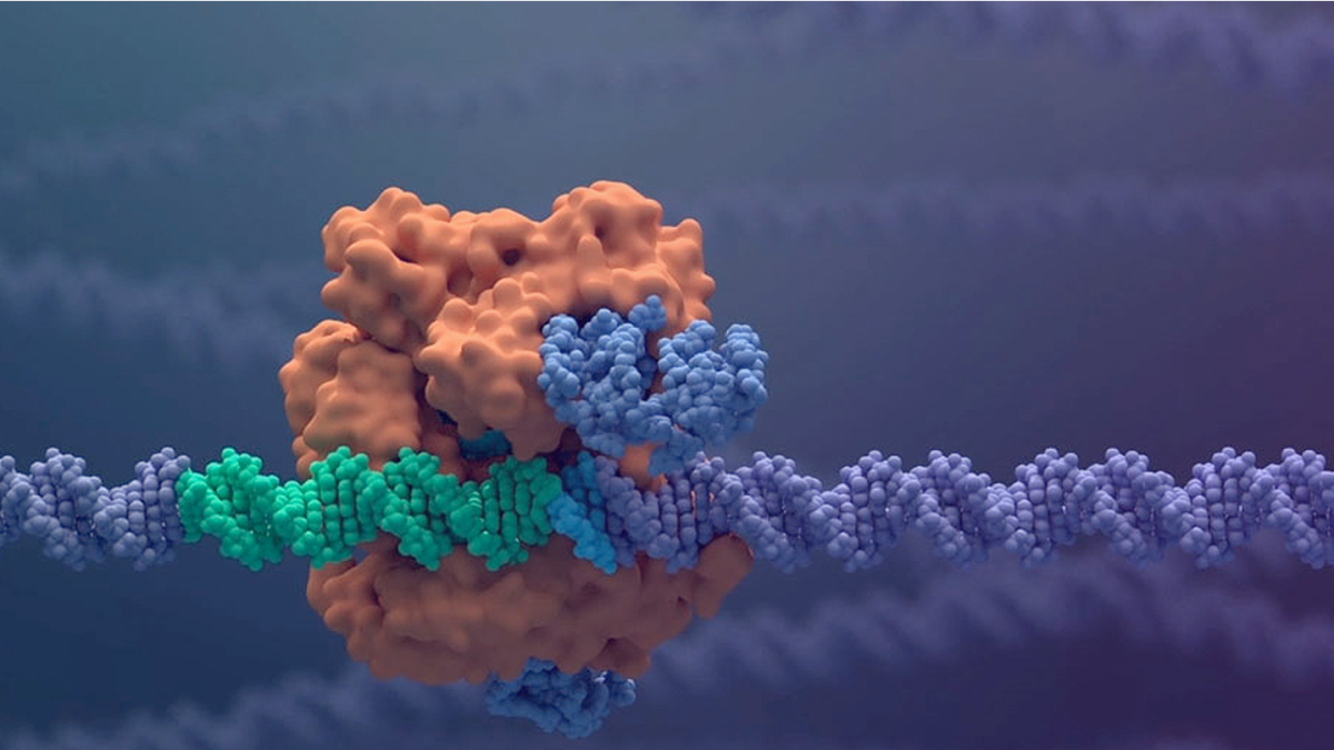
Exploring the human embryo with artificial intelligence. Objective: to better evaluate its characteristics before implantation. A team of experts combined 5 image analysis modules together using computer vision and, according to a study presented in Milan during the 38th annual meeting of the European Society of Human Reproduction and Embryology (Eshre), obtained an accuracy of 90 % in the prediction of chromosomally normal embryos. The method was developed by Ivi, an international company specializing in assisted reproduction. And 2,500 embryos have been analyzed.
According to the results illustrated in the Lombard capital, the percentage of accuracy is “close to that obtained from the conventional study on the embryo”, but the technique is “less invasive”. The study, which aims to improve more and more the practice of choosing the embryo to be implanted in the process of assisted procreation, was led by Marcos Meseguer, embryologist and scientific supervisor of Ivi Valencia. “Being able to assess the embryo’s implantation potential in this way allows us to improve the efficiency of a fundamental process in assisted reproduction, such as embryo culture and selection,” explained Francesco Gebbia, gynecologist, specialist in reproductive medicine.
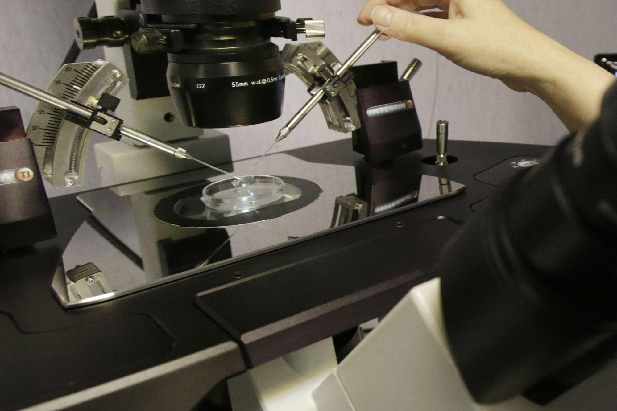
The new method consists in the reprocessing of data through complex algorithms that avoid having to manipulate the embryo, perform biopsies and extract cells, “thus obtaining a high capacity for success in identifying viable embryos to be transferred into the mother’s uterus”. The experts explained that these percentages “were previously only obtained with very invasive methods”. The technique used in the research, on the other hand, is “non-invasive” and is “universal, standardized and automatic” and would improve current methods of embryo evaluation.
“The focus of this study – added Daniela Galliano, director of the Pma Clinic in Rome, specialist in gynecology, obstetrics and reproductive medicine – responds to an indisputable reality: embryonic development does not occur in the same way in euploid embryos (chromosomally normal). and aneuploid (chromosomally abnormal). At this point, could Artificial Intelligence predict ploidy? The 5 modules we were able to analyze and combine show us that yes, it would be possible and reliable “.
What are the aspects studied? First, the morphokinetic parameters, a module that refers to the moments in which the most important events of embryonic development occur, that is, when the embryo divides into cells until it reaches the blastocyst stage. What the experts have verified is that, if an embryo arrives later in an event, than a euploid embryo taken as a reference, “its probability of being aneuploid increases considerably,” explained Gebbia. The second aspect is embryonic morphology: the automated study of this parameter shows that embryos with good morphology are more likely to be chromosomally normal. Morphology itself has a 68% predictive capacity for aneuploidy.
Third point: cellular activity: This module consists in measuring the diameter of a cell and the sum of all the diameters of the embryo’s cells at a specific time of its development (from 2 to 8 cells). “This automatically calculates the values which are then analyzed for 160 images, resulting in chromosomally abnormal or aneuploid embryos with a larger diameter length.” This is because they take longer to divide, the split produces a lot of movements and therefore increases the measurement, “continued Gebbia.
The fourth module then concerns the mitochondrial activity: it is a question of associating the smallest dimension that can be analyzed as regards the image, which is a pixel, with the size of a mitochondrion. Aneuploid embryos have a different number of pixels than euploid embryos, so this module helps predict aneuploidy with 77% accuracy. Last form: deformation / shrinkage: shrinkage occurs in approximately 20% of embryos. After automatically analyzing this event, it is observed that it occurs more frequently in aneuploid embryos.
“In short, artificial vision allows us to emulate the capabilities of our eyes on computers – concluded Gebbia – In other words, it aims to acquire, process, analyze and understand images of the real world in order to produce numerical or symbolic information that can be processed by a computer. And this ultimately allows us to verify that embryos behave differently during their development based on their chromosomal content and thus optimize the process of studying embryos and evaluating normal and viable embryos for the transfer”. The strategy developed by the scientists uses a complex algorithm developed by Ivi Valencia in collaboration with the Israeli company Aivf.
Experts believe that it can have an impact on increasing gestation rates, providing an objective and reliable forecast using a fast and inexpensive technique. “The international setting of the Milan Congress – commented Galliano – was also an opportunity for a constructive confrontation for a single goal: to look to the past to trace new paths for the future of medically assisted procreation and towards increasingly advanced techniques , in order to help fulfill the desire for parenthood of the many couples who for various reasons are forced to delay their journey “.
#Artificial #intelligence #study #embryo #Forecasts #accurate