Chemotherapy kills cancer cells. But the way these cells die appears to be different than previously expected. Researchers at the Netherlands Cancer Institute, led by Thijn Brummelkamp, have discovered a completely new way in which cancer cells die: due to the Schlafen11 gene.
The study was published on Science.
The pathway to cell death during chemotherapy
“This is a truly unexpected discovery. Cancer patients have been treated with chemotherapy for nearly a century, but this pathway to cell death has never been observed before. Where and when this occurs in patients will need to be further studied. This discovery could ultimately have implications for the treatment of cancer patients.”
Many cancer treatments damage cellular DNA. After too much irreparable damage, cells can begin their own death. High school biology teaches us that the p53 protein takes care of this process. p53 ensures repair of damaged DNA, but initiates cell suicide when the damage becomes too severe. This prevents uncontrolled cell division and cancer formation.
It seems like an infallible system, but the reality is more complex. “In more than half of tumors, p53 no longer works,” says Brummelkamp. “The key player p53 plays no role in this case. So why do tumor cells without p53 die anyway when you damage their DNA with chemotherapy or radiation? To my surprise, this turned out to be an unanswered question.”
His research group then discovered, together with colleague Reuven Agami’s group, a previously unknown way in which cells die after DNA damage. In the laboratory they administered chemotherapy to cells whose DNA they carefully modified. Brummelkamp says: “We were looking for a genetic change that would allow cells to survive chemotherapy. Our group has a lot of experience in selectively disabling genes, which we could apply perfectly here.”
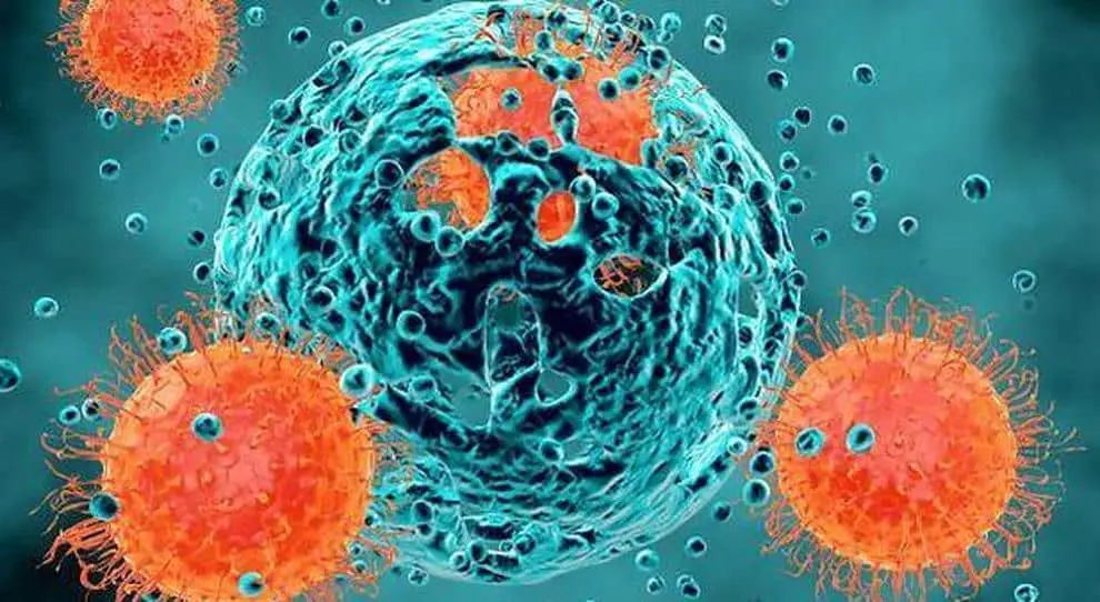
By turning off the genes, the research team discovered a new pathway to cell death driven by the Schlafen11 (SLFN11) gene. Lead researcher Nicolaas Boon said: “In case of DNA damage, SLFN11 switches off the cells’ protein factories: the ribosomes. This causes immense stress in these cells, leading to their death. The new pathway we discovered completely bypasses p53.”
The SLFN11 gene is not unknown in cancer research. It is often inactive in tumors of patients who don’t respond to chemotherapy, Brummelkamp says. “Now we can explain this connection. When cells lack SLFN11 they will not die in this way in response to DNA damage. The cells will survive and the cancer will persist.”
“This discovery opens up many new research questions, as usually happens in fundamental research,” says Brummelkamp.
“We have demonstrated our discovery in laboratory-grown tumor cells, but many important questions remain: where and when does this pathway occur in patients? How does it affect immunotherapy or chemotherapy? Does it affect the side effects of cancer therapy?
If this form of cell death proves to play a significant role in patients as well, this finding will have implications for cancer treatments. These are important questions to investigate further.
People have thousands of genes, many of which have functions that are unclear to us. To determine the role of our genes, researcher Brummelkamp developed a method using haploid cells.
These cells contain only one copy of each gene, unlike normal cells in our body which contain two copies.
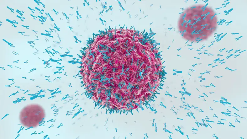
Managing two copies can be difficult in genetic experiments, because changes (mutations) often occur in only one of them. This makes it difficult to observe the effects of these mutations.
Together with other researchers, Brummelkamp has been uncovering crucial processes in diseases for years using this versatile method. For example, her group recently discovered that cells can produce lipids in a different way than previously known.
They have discovered how some viruses, including the deadly Ebola virus, manage to enter human cells. They delved into the resistance of tumor cells against specific therapies and identified proteins that act as brakes on the immune system, which is relevant for cancer immunotherapy.
In recent years, his team has discovered two enzymes that have remained elusive for four decades and have proven vital for muscle function and brain development.
The protein that controls resistance to chemotherapy
Despite the recent development of new targeted therapies, chemotherapies remain the most frequently used treatment to treat patients with advanced-stage cancers. Resistance to chemotherapy is a major cause of treatment failure and death in cancer patients.
It has been suggested that epithelial-mesenchymal transition (EMT), a process by which epithelial cells detach from neighboring cells and acquire invasive properties, plays a role in the acquisition of resistance to cancer therapy. However, the mechanism by which tumor cells exhibiting EMT resist anticancer therapy is currently unknown.
In a study published in Nature, researchers led by Prof. Cédric Blanpain, MD/Ph.D., WELBIO researcher, director of the Laboratory of Stem Cells and Cancer and professor at the Université Libre de Bruxelles, discovered that a protein called RHOJ enables cancer cells exhibiting EMT to resist cancer treatments by stimulating the repair of DNA damage caused by chemotherapy.
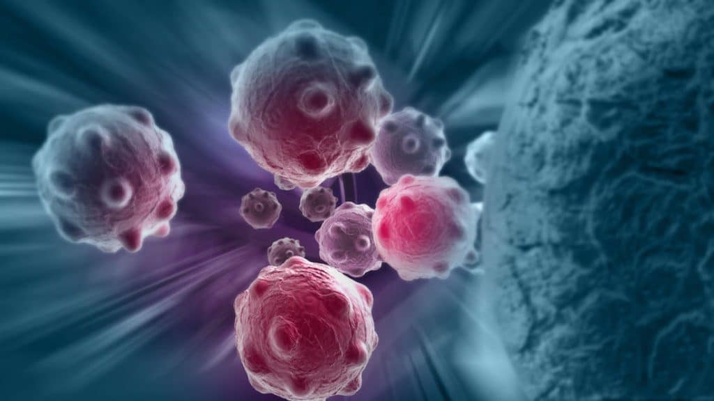
Maud Debaugnies and her colleagues have shown that tumor cells exhibiting EMT become resistant to chemotherapy treatment. They found that RHOJ expression was particularly high in chemotherapy-resistant cells. They then showed that by silencing RHOJ, tumor cells became sensitive to chemotherapy.
“It was particularly exciting to understand the mechanisms that allow tumor cells to resist chemotherapy, paving the way for the development of new and more effective therapeutic strategies to treat cancer,” says Maud Debaugnies, the first author of this study.
Maud Debaugnies and her colleagues then studied which RHOJ mechanisms make tumor cells resistant to chemotherapy. Chemotherapy induces DNA damage in tumor cells which triggers the death of these cells. They found that RHOJ can activate the chemotherapy-induced DNA damage repair pathway, allowing cancer cells to repair DNA lesions and escape cell death.
“Our discovery that inhibition of a single gene can make tumor cells sensitive to chemotherapy opens new avenues for the development of RHOJ-targeted drugs that should decrease chemotherapy resistance in patients with tumors exhibiting EMT,” says Prof Cedric Blanpain, the director of this studio.
Selective protection of normal cells from chemotherapy, while killing drug-resistant tumor cells
Cancer therapy is limited by toxicity in normal cells and drug resistance in tumor cells. In his latest review, Mikhail V. Blagosklonny, M.D., Ph.D., of Roswell Park Comprehensive Cancer Center, discusses the theory that cancer resistance to certain therapies can be exploited for the protection of normal cells, while simultaneously allowing for Selective killing of resistant cancer cells using antagonistic drug combinations, which include cytotoxic and protective drugs.
“No cancer cell, no matter how resistant, can survive chemotherapy in a cell culture. In the body, however, cancer therapy is limited by killing or damaging normal cells.
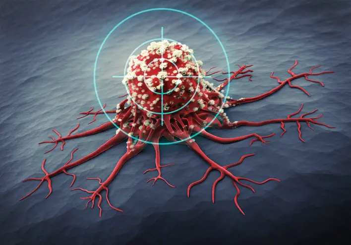
Selective protection of normal cells from chemotherapy would increase the therapeutic window, improving therapeutic outcome. It goes without saying that reduced side effects and improved quality of life are very important for a cancer patient,” says Dr. Blagosklonny.
Depending on the mechanisms of drug resistance in tumor cells, protection of normal cells can be achieved with inhibitors of CDK4/6, caspase, Mdm2, mTOR and mitogen kinases. When normal cells are protected, the selectivity and potency of multidrug combinations can be further improved by adding synergistic drugs, in theory eliminating the deadliest tumor clones with minimal side effects.
“I also talk about how the recent success of Trilaciclib can promote similar approaches in clinical practice, how to mitigate systemic side effects of chemotherapy in patients with brain tumors, and how to ensure that protective drugs protect only normal cells (not tumor cells). ) in a particular patient,” adds Dr. Blagosklonny.
A blood sample 24 hours after starting chemotherapy can predict survival
Researchers at the University of Bergen in Norway have found a new method that can predict within hours whether or not some cancer patients will survive after chemotherapy.
Acute myeloid leukemia is an aggressive blood cancer with poor survival. Although initial response rates to chemotherapy are high, patients often relapse due to the selection and development of chemotherapy-resistant leukemic cells.
“When treating patients with leukemia, it is difficult to quickly follow whether the patient responds to therapy or not,” says Benedicte Sjo Tislevoll, a researcher at the University of Bergen and leader of the new study.
The response to therapy is currently measured after weeks or months of treatment, thus wasting important time. However, an immediate response to chemotherapy can be measured by studying the functional properties of leukemia cells.
“Our results show that ERK1/2 protein increases within the first 24 hours after chemotherapy in patients who have a poor response to therapy. We believe that this protein is responsible for the resistance of tumor cells to chemotherapy and can be used to distinguish responders from non-responders,” says the researcher.
“We believe this is an important key to our understanding of cancer, and our goal is to use this information to modify treatment early for patients who do not respond to therapy,” concludes Tislevoll.
#path #tumor #cell #death #due #chemotherapy #discovered
