The most common reason for miscarriages are chromosomal defects which can be difficult to diagnose. To try to find a solution to this problem, a research team fromUniversity of Copenhagen, in a recent study he developed a new diagnostic method that can clarify the appearance of chromosomal defects and the chromosomal changes associated with a disease and how to support the diagnosis.

The results of the Research have been published in the scientific journal Nature.
Chromosomal defects: here’s how to prevent the consequences
In Denmark, fertility problems are becoming more and more frequent. The most common cause of miscarriage are chromosomal defects: in more than half of the miscarriages that occur during the first 12 weeks, chromosomal defects are found in the fetus.
Often doctors have no idea which chromosomal defect is involved in miscarriage, and it is precisely for this reason that parents themselves have the tools to know what to do to bring the pregnancy to term. However, New research from the University of Copenhagen may help solve this age-old problem.
In fact, a newly developed technology can characterize chromosomes with an unprecedented level of detail and can help discover new chromosomal errors, which cannot be diagnosed with current methods.: “The dream is that we can take a chromosome sample from a person who has a fertility problem, for example, and then use our method to analyze chromosomes and determine if something is wrong. It could potentially also be used to investigate other chromosomal defects and diseases, such as cancer “, said Professor Ian Hickson of the Department of Cellular and Molecular Medicine, who led the research.
Today it is already normal practice in Danish hospitals to examine chromosomes for defects. But the new method will allow to study the chromosomes carefully in more depth: “Today, the chromosomes examined are exposed to chemicals that repair them “explained Professor Hickson.
“We use optical tweezers and a super-resolution microscope to examine the chromosomes as if they were inside cells, where they are flexible and mobile. With that method, we can move, push and pull the chromosomes to see if there are any abnormalities“, Ian Hickson specified. With a super-resolution microscope, they can see in detail what happens to the chromosome when they manipulate it: “We can detect multiple hidden chromosomal defects. Perhaps the chromosome, for example, falls apart when we pull one end. So we know we are dealing with a chromosomal defect. But you can’t know until you’ve stretched the chromosome“, Continued the expert.
“With this method we can obtain a precise calculation of the mechanical properties of chromosomes and this can provide a detailed view of the underlying structure. If a diseased chromosome has a weaker structure or a weak spot we will be able to measure it with our method“, He intervened Christian Friberg Nielsenone of the main authors of the research.
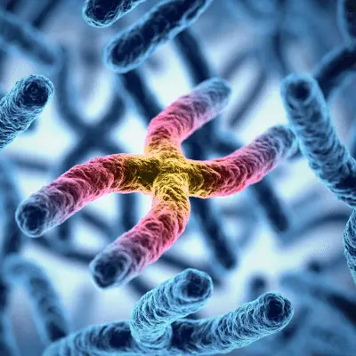
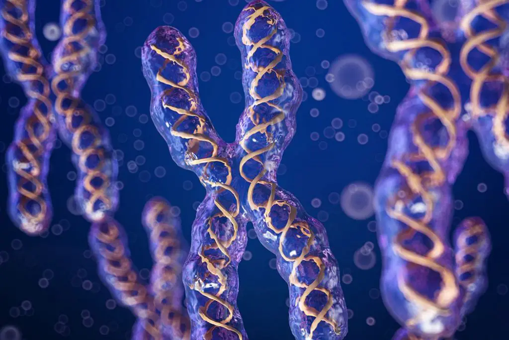
To find out what a faulty chromosome looks like, scientists first had to study several hundred healthy chromosomes: “A chromosome is a remarkable structure. It consists of two DNA molecules. The DNA in a single cell’s chromosomes is two meters long but is encased within a tiny cell with a diameter less than the thickness of a human hair. The two meters of DNA must be packed and folded until the chromosomes adopt the characteristic ax shape, and only then can we see the individual chromosomes using a microscope“, Hickson specified.
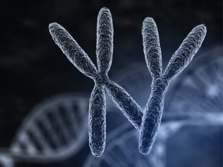
Ian Hickson pointed out that there is a very fine balance with chromosomes and it doesn’t take more than a small chromosomal error before it has major consequences: “The best known genetic chromosomal abnormality is Down syndrome, which is caused by a baby being born with three copies of chromosome 21 instead of two. While a lesser known defect, in which the embryo has three copies of chromosome 16, is the most common cause of miscarriage. It shows how delicately balanced the system is“, Added the scientist.
About 1% of the world population lives with a chromosomal defect. Some with chromosomal defects can live normal lives, such as men with double Y syndrome who have two copies of the male sex chromosome. For other individuals with chromosomal defects it becomes much more disabling and can lead to premature death.
The research team at the University of Copenhagen therefore believes that the new method can be used to diagnose chromosomal defects and thus become more aware of how to screen for chromosomal defects in fetuses. In the long run, it may also be possible to help people living with a chromosomal defect, for example by screening their chromosomes to suggest treatments that compensate for some features of the defect.
In these cases, the hope is that the new method can help determine if the chromosomes are actually hiding the cancer-causing changes. Ian Hickson estimated the method will likely be tested in the lab for five to ten years before researchers can try it in clinical trials.
#Chromosomal #defects #diagnostic #method #arrives


