New research onAlzheimer’s has shed light on the complex interaction between cellular proteins and how they impact neurons in neurodevelopmental disorders and disease.
The study conducted by the University of Exeter and published on Open Biology discovered the key role that the Contactin-4 protein (encoded by the CNTN4 gene) plays in the formation of neurons.
Researchers began studying CNTN4 because its role in autism was known, but its functional roles were not well understood. The team explored how CNTN4 works within the brain, particularly its interactions with proteins involved in neurodegenerative diseases such as Alzheimer’s disease.
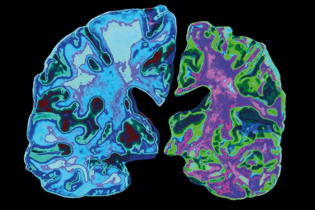
For the first time, researchers studied mice that had the CNTN4 gene deleted in the cortex, the brain region responsible for key functions including memory, thinking and reasoning. They found that neurons developed differently in the cortex region.
Researchers have demonstrated for the first time in human cells the interaction between the CNTN4 genes and APP, a gene strongly linked to Alzheimer’s disease, revealing a co-dependent relationship essential for brain development, and in particular for growth healthy neurons.
The investigators found that CNTN4 not only contributes to neural elongation in the frontal cortex region of the brain, but also that CNTN4 expression is regulated through a relationship with APP.

Using studies in genetically modified human cells, the team also found that there is a complex interaction between CNTN4 and APP. If CNTN4 is deleted, APP levels decrease, but not to zero. Scientists believe that APP can compensate for the loss of CNTN4 and vice versa.
Lead author of the study, Dr Rosemary Bamford from the University of Exeter Medical School, said: “It was truly remarkable to discover that CNTN4, a gene linked to developmental processes, also plays a role in modulating factors involved in Alzheimer’s disease. Alzheimer’s. This intersection of developmental and neurodegenerative pathways offers exciting new insights into the broader implications of these proteins.”
Senior author Dr Asami Oguro-Ando, from the University of Exeter Medical School, said: “Looking ahead, my group is keen to further analyze the molecular mechanisms underlying the interaction between CNTN4 and APP and explore the their broader implications for disorders such as Alzheimer’s and Alzheimer’s.” autism spectrum disorder. Our next steps involve elucidating how the CNTN4-APP interaction affects neural activity.
Understanding this interaction is crucial as it represents a fundamental step towards a comprehensive understanding of neurodevelopmental and neurodegenerative disorders.
The “MUSIC map” reveals that some brain cells age faster and are more prevalent in Alzheimer’s
Engineers at the University of California at San Diego have found that some brain cells age more rapidly than others and are disproportionately abundant in individuals with Alzheimer’s disease.
Furthermore, the researchers observed sex-specific differences in the aging process of some brain cells, with the female cortex showing a higher ratio of “old” oligodendrocytes to “old” neurons than the male cortex.
The discoveries were made possible by a new technique called MUSIC (multinucleic acid interaction mapping in single cells), which allows researchers to peek inside individual brain cells and map the interactions between chromatin – which is the tightly coiled form of DNA – and RNA.
This technique allows researchers to visualize these interactions at single-cell resolution, as well as study how they affect gene expression.
The work is detailed in a paper published in Nature.
“MUSIC is a powerful tool that may allow us to dig deeper into the complexities of Alzheimer’s disease,” said senior study author Sheng Zhong, professor in the Shu Chien-Gene Lay Department of Bioengineering at the Jacobs School of UC San Diego Engineering.
“The technology has the potential to help us uncover new molecular mechanisms underlying Alzheimer’s pathology, which could pave the way for more targeted therapeutic interventions and better patient outcomes.”

The human brain is home to a complex network of cells that communicate and interact in complex ways. Within each of these cells lies a dynamic interplay of genetic components, including chromatin and RNA, that dictate crucial cellular functions. As brain cells grow and age, these interactions between chromatin and RNA change.
And within each cell these complexes can vary widely, especially in mature cells. However, unraveling the nuances of these interactions has remained a formidable challenge.
Enter MUSIC, a cutting-edge tool that offers a window into the inner workings of individual brain cells. Using MUSIC, Zhong’s team analyzed postmortem brain samples, specifically human frontal cortex tissue, obtained from 14 donors aged 59 or older, some with Alzheimer’s disease and some without.
They found that different types of brain cells showed distinct patterns of interactions between chromatin and RNA. Interestingly, cells with fewer short-range chromatin interactions tended to show signs of aging and Alzheimer’s disease.
“With this transformative single-cell technology, we found that some brain cells are ‘older’ than others,” Zhong said. Notably, individuals with Alzheimer’s disease had a higher percentage of these older brain cells than healthy individuals, she explained.
Researchers say the discovery could help in the development of new treatments for Alzheimer’s disease.
“If we could identify dysregulated genes in these aged cells and understand their functions in local chromatin structure, we could also identify new potential therapeutic targets,” said study co-author Xingzhao Wen, a PhD in bioinformatics. candidate in Zhong’s lab.
The study also discovered sex-specific differences in the aging of brain cells. In the cortex of female mice, the researchers found a higher ratio of aged oligodendrocytes to aged neurons. Oligodendrocytes are a type of brain cell that provide a protective layer around neurons. Given their critical role in maintaining normal brain function, an increased prevalence of aged oligodendrocytes could potentially exacerbate cognitive decline.
“The disproportionate presence of old oligodendrocytes in the female cortex could shed new light on the increased risks of neurodegenerative and mental disorders observed in women,” Wen said.
Next, researchers will work to further optimize MUSIC so that they can use it to identify factors – such as regulatory genes and genetic circuits – responsible for the accelerated aging observed in specific brain cells.
“Next, we will devise strategies to prevent the activity of these genes or circuits, in the hope of mitigating brain aging,” Zhong said.
Gene involved in neuronal vulnerability in Alzheimer’s disease identified
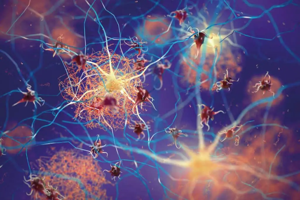
The early stages of neurodegenerative disorders are characterized by the accumulation of proteins in distinct populations of brain cells and the degeneration of these cells.
For most diseases, this pattern of selective vulnerability is unexplained, but could provide important insights into disease mechanisms.
Alzheimer’s disease (AD), the leading cause of dementia worldwide, is defined by the appearance of two characteristic pathological lesions, amyloid plaques (extracellular aggregates of Aβ peptides) and neurofibrillary tangles (intracellular aggregates of hyperphosphorylated tau or NFT). While plaques are widespread in the neocortex and hippocampus, NFTs follow a well-defined regional pattern starting in principal neurons of the entorhinal cortex.
In a new study from Boston University’s Chobanian & Avedisian School of Medicine, researchers have identified a gene that they believe may lead to the degeneration of neurons most vulnerable to AD.
“We are trying to understand why some neurons in the brain are particularly vulnerable during the early stages of AD. Why they accumulate and degenerate very early is not known.
We believe that elucidating this vulnerability would enable a new therapeutic avenue for AD,” the correspondent said. author Jean-Pierre Roussarie, Ph.D., assistant professor of anatomy and neurobiology at the school.
In collaboration with leading computational genomics experts at Rice University, the BU researchers, along with co-corresponding author Patricia Rodriguez-Rodriguez, Ph.D., of the Karolinska Institute, used cutting-edge analysis tools with machine learning to identify the DEK gene as perhaps responsible for the vulnerability of neurons in the entorhinal cortex.
They injected viruses into the entorhinal cortex of experimental models and laboratory-grown neurons to manipulate DEK gene levels. When they reduced levels of the DEK gene, the vulnerable neurons began to accumulate tau and degenerate.
According to the researchers, preventing the degeneration of these neurons by targeting DEK or proteins that work with DEK would prevent patients from developing memory loss and reduce the disease before it spreads to larger areas of the brain. “Given that neurons in the entorhinal cortex are necessary for the formation of new memories, and because they are so vulnerable and the first to die, this explains why the first symptom of AD is the inability to form new memories,” Roussarie said.
The researchers believe these findings are the first step in understanding how these fragile neurons die, but they hope to discover additional genes to fully understand what leads to the death of critical memory-forming neurons.
#Alzheimerslinked #proteins #affect #neuronal #growth
